[10000印刷√] e coli under microscope gram stain 153380-What gram stain is e coli
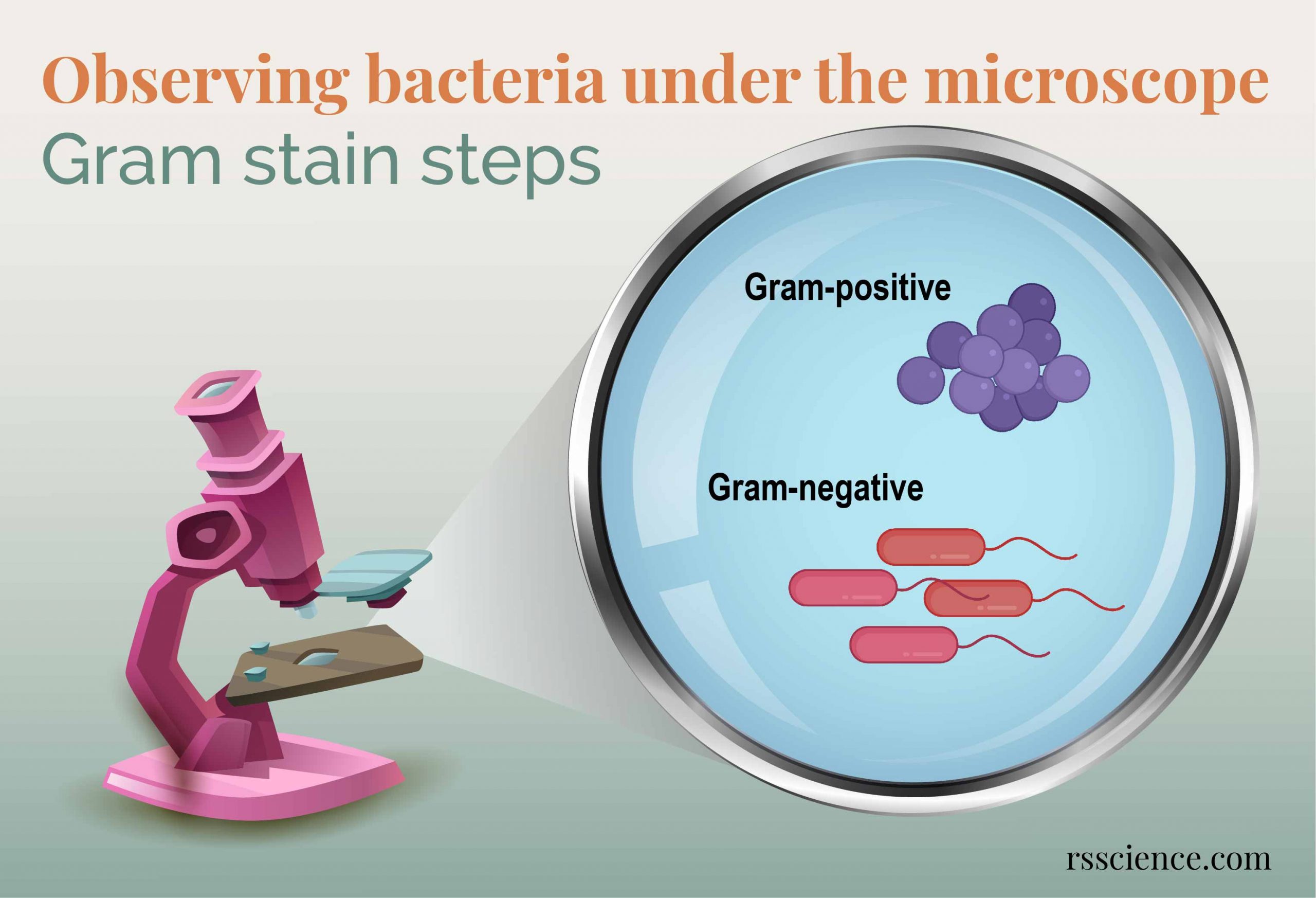
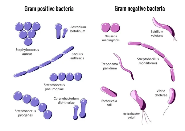
Gramnegative bacteria have an outer cell membrane while grampositive bacteria don't A simple test with a violet color dye (called gram stain) is used to determine the type of the bacteria when looked at under a microscope The bacteria with a membrane repel the gram stain and remain pink, and therefore are called gramnegativeLastly, a drop of oil was added to the slide so the sample could be viewed under the microscope using the oil immersion lens Results The E coli had a Gram Stain reaction color of pink and classified as Gramnegative Both the S epidermidis and B subtilis had a Gram Stain reaction color of purple and then classified as Grampositive (Table 1)Gram stain allows you to classify into Gramnegative rods (which includes E coli) and Grampositive rods (which include Bacillus, Corynebacterium, and some others)
Http Coltonanderson1 Weebly Com Uploads 2 4 3 0 Manual Pdf
What gram stain is e coli
What gram stain is e coli-A)All cells appear colorless B)Most cells would appear blue C)Most cells would appearA)All cells appear colorless B)Most cells would appear blue C)Most cells would appear


Pathogenic E Coli
Gram Staining of Micrococcus luteus,Escherichia coli, and Serratia marcescens INTRODUCTION In 14, a Danish botanist named Hans Christian Gram developed the popular staining technique known as the "gram stain"His background in studying plants at the University of Copenhagen introduced him to observing different tissues under a microscope, and thus propelled his interest in pharmacologyAnswer to Can you check this lab report Please then tell me what I need to fixE coli bacteria are gramnegative, so they stain pink in a gram test E coli bacteria were first identified by bacteriologist Dr Escherich in 15 Since then, more than 700 different serotypes have been identified Some of these strains are pathogenic, but most are not A strain known as uropathogenic E coli, or UPEC, is known to cause
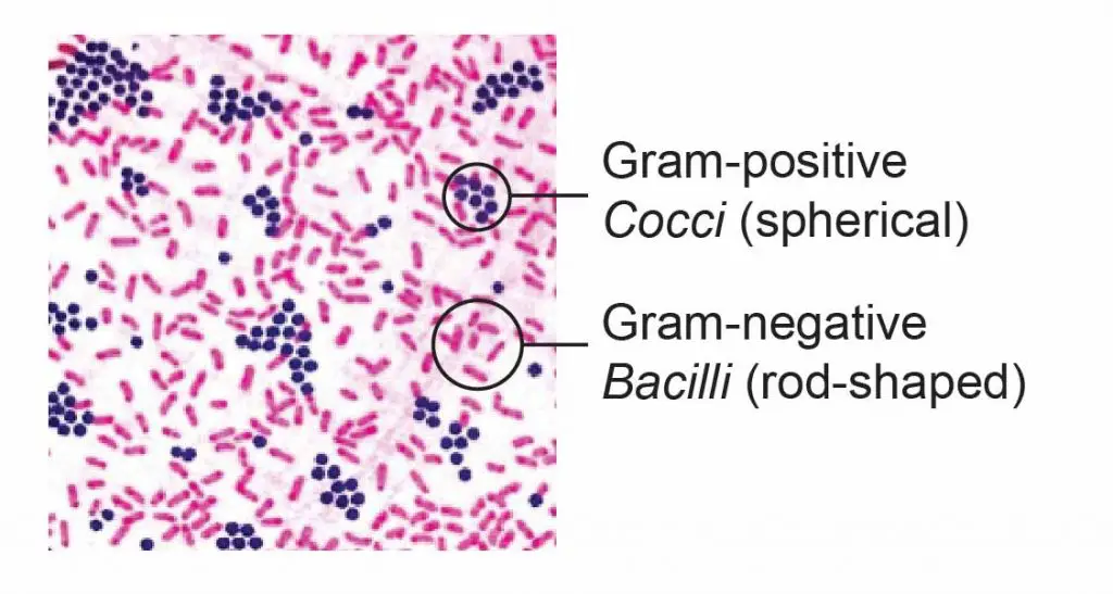
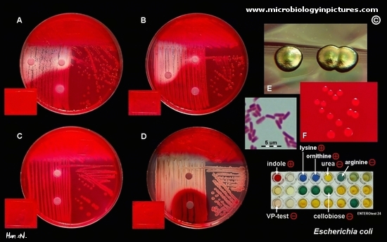
Gramstain Grampositive cocci and Gramnegative bacilli Microscopic appearance Cocci inSalmonella includes a group of gramnegative bacillus bacteria that causes food poisoning and the consequent infection of the intestinal tract While some of the infections can be easily treated, some of the strains have been shown to resist antibiotic treatmentOn Endo agar it looks like lactose negative)All four strains are mannitol positive (best seen in fig D), cellobiose negative (strains A, B)
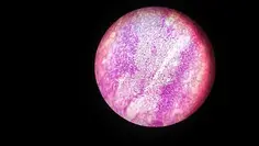
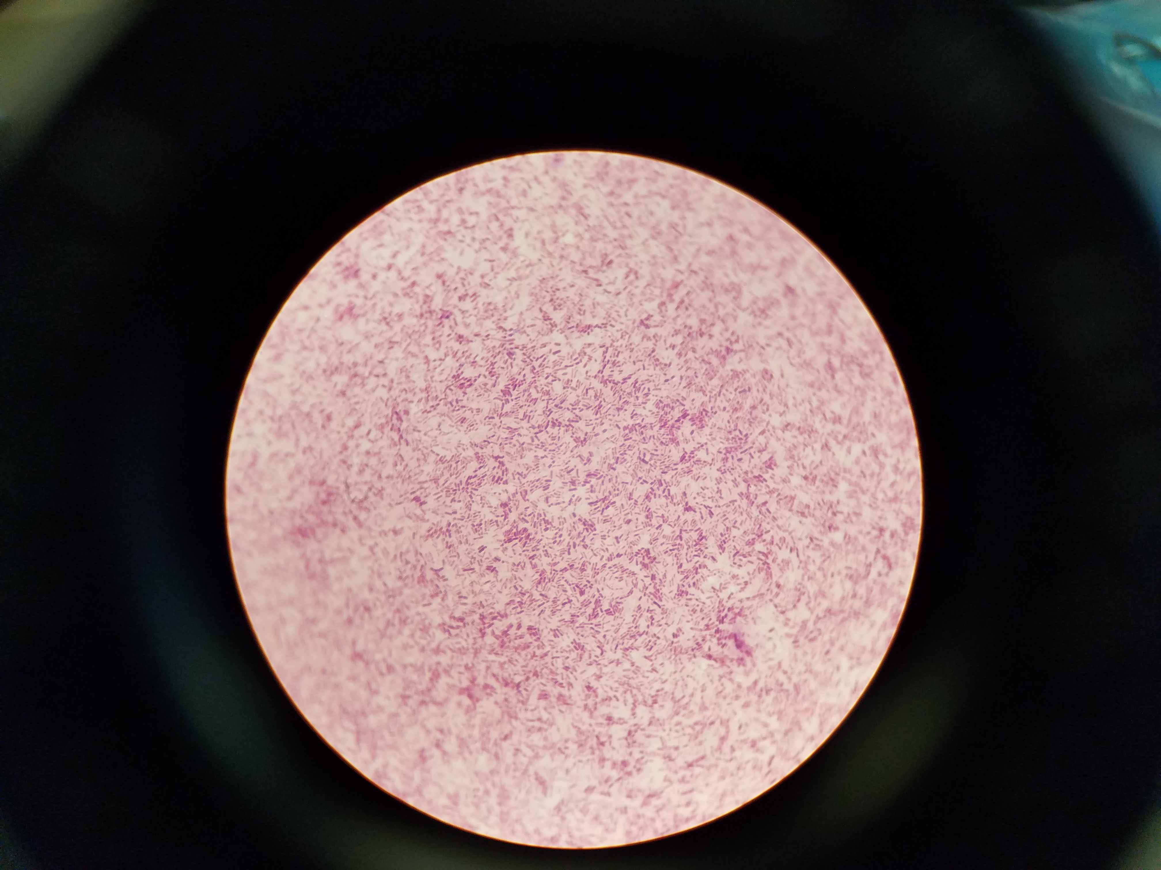
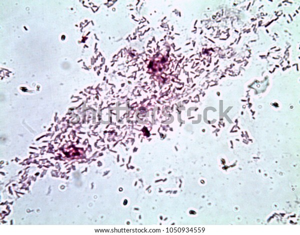
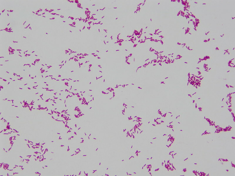
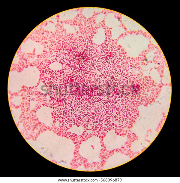
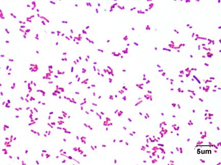
List the reagents of the gram stain technique in order and their general role in the staining process 1 Crystal violet primary stain 2 Grams iodine mordant 3 Alcohol decolorizer Escherichia coli under the microscope if you forgot to apply safranin colorless Escherichia coli under the microscope if you forgot to apply decolorizerThe bacterium turned purple when Gram stained indicating a Gram positive microbe The bacterium showed single and clustered rods when observed under the compound microscope at 1000X magnification as Bacillus subtilis Gram Mixed Culture (Gram Stain) E coli and S epidermidis (1000X) Klebsiella pneumoniae (1000X) Capsule StainE coli is a Gramnegative rodshaped bacteria When Gram stained, the organism looks pink or red Here are a couple of pictures of a Gram stain of E coli that I did under the 100X objective lens on a standard light microscope You can of course


Escherichia Coli Bacteria E Coli Stock Footage Video 100 Royalty Free Shutterstock


Gram Stain Images Microbiology Stain Study Tools
Finally place slide under the microscope to view results Results When looking through the microscope it is apparent the gram stain has both gram positive Staph Aureus and gram negative E coli bacteria Again E coli is negative because it is pink in color, you can also see it's rod shaped StaphE coli is an intestinal organism, and staphylococcus epidermidis resides on the skin The differential stage of the Gramstain is the application of A)Iodine B)Crystal violet what color would the cells likely be when viewed under the microscope?Escherichia coli Four different strains of Escherichia coli on Endo agar with biochemical slope Glucose fermentation with gas production, urea and H 2 S negative, lactose positive (with exception of strain D "late lactose fermenter";
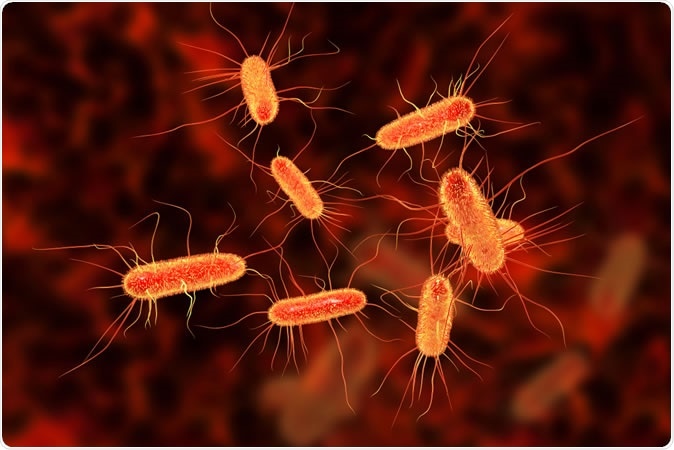


E Coli Infections Linked To Chopped Salad Kits Cdc Warns


Firmicutes Wikipedia
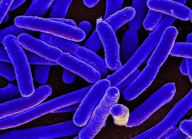
E coli under the microscope Escherichia coli (E coli) is a bacterium commonly found in various ecosystems like land and water Most of the strains of E coli are harmless, but some strains are known to cause diarrhea and even UTIs E coli is commonly studied as they are considered as a standard for the study of different bacteriaThe technician decides to make a Gram stain of the specimen This technique is commonly used as an early step in identifying pathogenic bacteria After completing the Gram stain procedure, the technician views the slide under the brightfield microscope and sees purple, grapelike clusters of spherical cells (Figure 4)Escherichia coli (/ ˌ ɛ ʃ ə ˈ r ɪ k i ə ˈ k oʊ l aɪ /), also known as E coli (/ ˌ iː ˈ k oʊ l aɪ /), is a Gramnegative, facultative anaerobic, rodshaped, coliform bacterium of the genus Escherichia that is commonly found in the lower intestine of warmblooded organisms (endotherms) Most E coli strains are harmless, but some serotypes (EPEC, ETEC etc) can cause serious food


Gram Stain
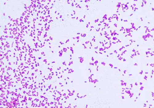


Gram Negative Bacteria Images Photos Of Escherichia Coli Salmonella Enterobacter
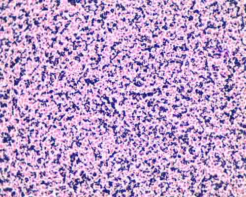
See more pathogens and parasites on our channel & please subscribe!Mixed Gram & Gram Bacteria Under Microscope (Click on image to enlarge) Gram stain mixed sample od Staphylococcus (Gram , purple cocci) & E coli , (Gram pink rods) @ 1000xTMGram stain e coli under microscope 100x › microscope e coli gram stain bacteria › microscope escherichia coli gram stain e coli Gram Stain E Coli Microscope Written By MacPride Tuesday, July 23, 19 Add Comment Edit Puzzlepix Figure 1 From Detection Of Multidrug Resistant And Shiga Toxin


Gram Negative Pink Colored Small Rod Shape E Coli Under Light Download Scientific Diagram


Photo Gallery Of Pathogenic Bacterial
A slide is also not acceptable for examination if microorganisms that should be grampositive appear pink This may indicate that either the Gram's iodine was not applied or the slide was overdecolorized Staining grampositive and gramnegative control slides along with the patient's smear would confirm that proper staining technique was usedThere are two type of step in Gram stain techniques which are threestep Gram stain and fourstep Gram stain However, most clinical laboratories are currently using the four step Gram stain The colour of the microorganisms that can be seen under the light microscope can be either purple or redpink with the background of the tissue is yellowThe purpose of this experiment was to determine the Gram stain of the certain slant cultures we used in class, Escherichia coli and Staphylococcus aureus, under a microscope Since it is the most important staining procedure in microbiology, it is used to distinguish between grampositive and gramnegative organisms


Biol 230 Lecture Guide Gram Stain Of A Mixture Of Gram Positive And Gram Negative Bacteria


What Are Examples Of Gram Negative Bacteria Quora
Procedure of Gram stain for E coli Cover the smear with crystal violet and allow it to stand for one minute Rinse the smear gently under tap water Cover the smear with Gram's iodine and allow it to stand for one minute Rinse smear again gently under tap water Decolorize the smear with 95% alcohol Rinse the smear again gently under tapGram Staining Escherichia Coliobserved Under Optical Stock Photo Eisco Prepared Microscope Slide Escherichia Coli Smear Gram Asmscience Examination Of Gram Stains Of Urine Gram Stain Tissue Trol Control Slides From Mouse Lung Tissue Gram Stain Of E Coli Bacterium A Gram Stain Of ShowsEscherichia coli Gram stained smear under microscopeRod shapedpink in colorthat's why Gram negative Bacilli#GramStain#GNB#GNR


Lm Of The Gram Negative Bacteria E Coli Stock Image B230 0001 Science Photo Library
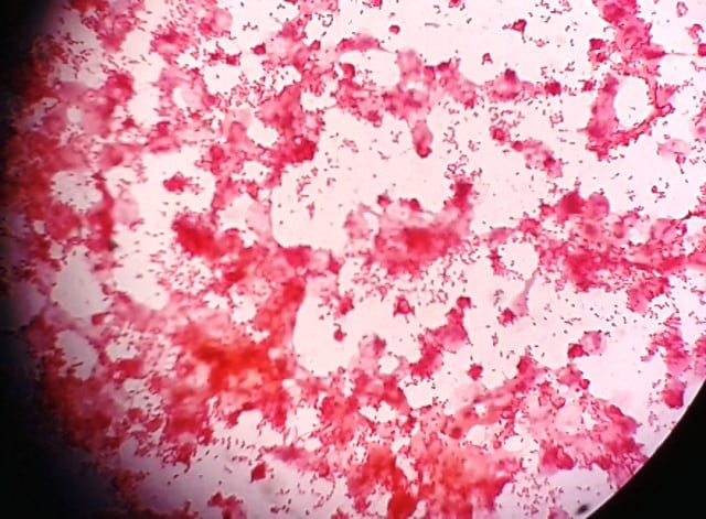


What Are Gram Negative Bacteria
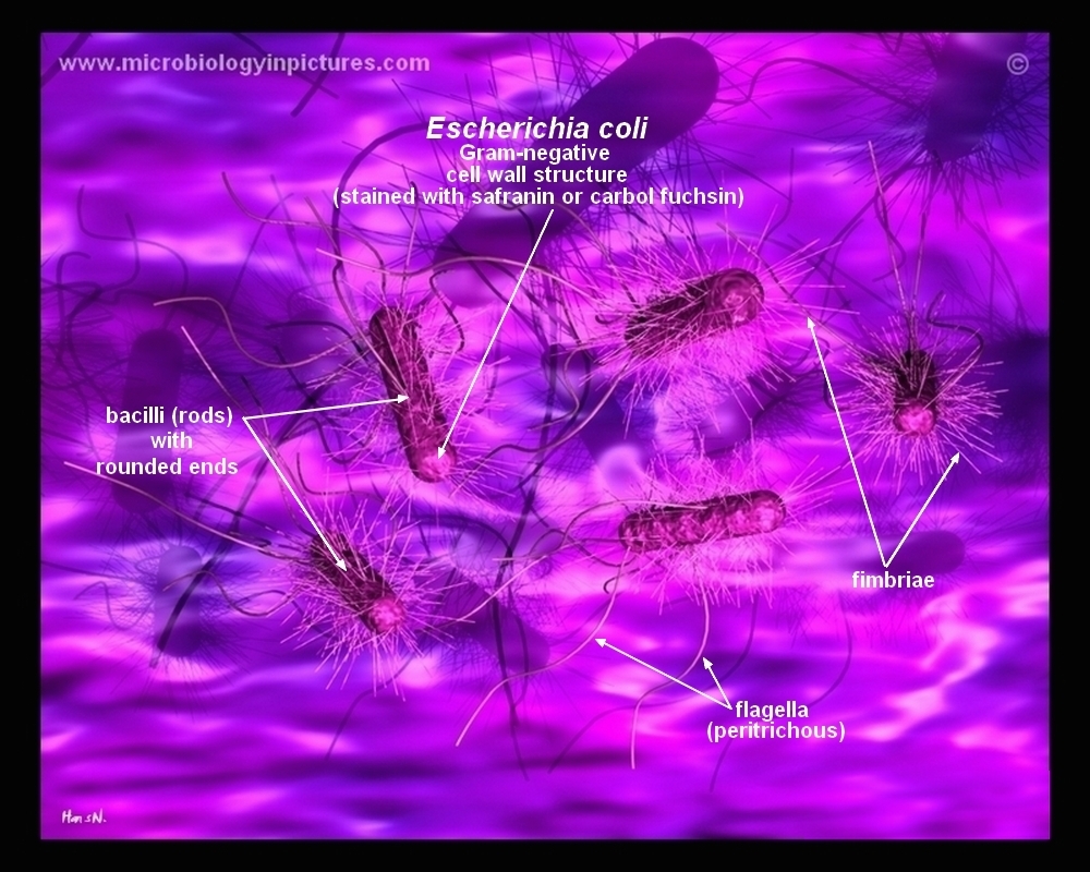
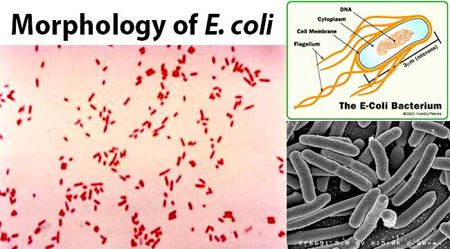
Ecoli is gram negative bacteria it give pink colour for staining bacteri with gram stain first make smear then put a drop of crstal violet on smear for one minute now wash it with distill waterExamples of Bacteria Under the Microscope Escherichia coli Escherichia coli (Ecoli) is a common gramnegative bacterial species that is often one of the first ones to be observed by students Most strains of Ecoli are harmless to humans, but some are pathogens and are responsible for gastrointestinal infections They are a bacillus shaped bacteria that has a very fast growth (they can double every minutes), which is one of the main reasons they are used in research2 Morphology and Staining of Escherichia Coli E coli is Gramnegative straight rod, 13 µ x 0407 µ, arranged singly or in pairs (Fig 281) It is motile by peritrichous flagellae, though some strains are nonmotile Spores are not formed Capsules and fimbriae are found in some strains 3 Cultural Characteristics of Escherichia Coli


Infection Landscapes Escherichia Coli
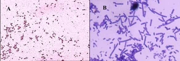


How These 26 Things Look Like Under The Microscope With Diagrams
On Endo agar it looks like lactose negative)All four strains are mannitol positive (best seen in fig D), cellobiose negative (strains A, B)E Coli Microscopy To determine whether a strain (s) is present in a sample, it's necessary to stain the sample Here, Gram stain is used as it helps distinguish between the gram positive and gram negative bacteria in a sample Being a differential stain, Gram stain is more complex compared to more simple stains like methylene blueFree YouTube Audio Library Music was used Carol of the Bells by Quincas Moreira


Escherichia Coli Slide W M Science Lab Microbiology Supplies Amazon Com Industrial Scientific
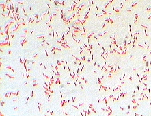


Photo Gallery Of Pathogenic Bacterial
Salmonella includes a group of gramnegative bacillus bacteria that causes food poisoning and the consequent infection of the intestinal tract While some of the infections can be easily treated, some of the strains have been shown to resist antibiotic treatmentMagnification 1000× Gram stain Result Gramnegative rods wwwbacteriainphotoscomView under Microscope (10x, add oil then 100X) Ecoli TSA 7 Heat slide and label 8 Preform Aseptic technique to retrieve bacteria from the slant and smear on slide (remember to add a drop of water) 9 Air Dry Slide 10 Heat Fix slide 11 Preform Gram Stain 12 View under Microscope (10x, add oil then 100X) Mixed TSA and TSB 13


How E Coli Bacteria Look Like


E Coli Under The Microscope Types Techniques Gram Stain Hanging Drop Method
On staining, E coli appear as nonsporeforming, Gramnegative rodshaped bacterium Routine urine cultures should be plated using calibrated loops for the semiquantitative method Note The most commonly used criterion for defining significant bacteriuria is the presence of ⩾10 5 CFU per milliliter of urineOn staining, E coli appear as nonsporeforming, Gramnegative rodshaped bacterium Routine urine cultures should be plated using calibrated loops for the semiquantitative method Note The most commonly used criterion for defining significant bacteriuria is the presence of ⩾10 5 CFU per milliliter of urineProcedure of Gram stain for E coli Cover the smear with crystal violet and allow it to stand for one minute Rinse the smear gently under tap water Cover the smear with Gram's iodine and allow it to stand for one minute Rinse smear again gently under tap water Decolorize the smear with 95% alcohol Rinse the smear again gently under tap


E Coli Gram Stain Mikrobiologiya


Mycolight Rapid Fluorescence Bacterial Gram Stain Kit t Bioquest
E coli is an intestinal organism, and staphylococcus epidermidis resides on the skin The differential stage of the Gramstain is the application of A)Iodine B)Crystal violet what color would the cells likely be when viewed under the microscope?Escherichia coli (/ ˌ ɛ ʃ ə ˈ r ɪ k i ə ˈ k oʊ l aɪ /), also known as E coli (/ ˌ iː ˈ k oʊ l aɪ /), is a Gramnegative, facultative anaerobic, rodshaped, coliform bacterium of the genus Escherichia that is commonly found in the lower intestine of warmblooded organisms (endotherms) Most E coli strains are harmless, but some serotypes (EPEC, ETEC etc) can cause serious food1 Escherichia coli a Fix a smear of Escherichia coli to the slide as follows 1First place a small piece of tape at one end of the slide and label it with the name of the bacterium you will be placing on that slide 2Using the dropper bottle of deionized water found in your staining rack, place 1/2 of a normal sized drop of water on a clean slide by touching the dropper to the slide


Photomicrographs


Q Tbn And9gcssskwic Hp Oxiadhcwlxxp0xfsk4jr3zus4de387pietcequq Usqp Cau
Answer to Can you check this lab report Please then tell me what I need to fixThe slide is stained with safranin for 1min, then rinse with water very gently until no color is coming from the water Blot dry with absorbent paper Place the slide on the microscope to view the sample The grampositive bacteria will stain dark blue or purple The gramnegative bacteria will stain pink In this figure A procedure of Gram stain1 E coli stained with crystal violet @ 100xTM It is not possible to see individual bacteria at this magnification, but viewing at 100XTM is a necessary step in focusing to scope to see at higher magnifications 2 E coli stained with crystal violet @ 1000xTM


Escherichia Coli Colony Morphology And Microscopic Appearance Basic Characteristic And Tests For Identification Of E Coli Bacteria Images Of Escherichia Coli Antibiotic Treatment Of E Coli Infections


Esbl Producing Enterobacterales Hai Cdc
Gram Staining Now that the slide has been heat fixed, you may now begin to stain the organism This is started by applying a few drops of crystal violet for 30 seconds, then rinsing with water for 5 seconds Next, cover the slide with Gram's Iodine and let sit for a full minute before rinsing with water for another 5 secondsThe technician decides to make a Gram stain of the specimen This technique is commonly used as an early step in identifying pathogenic bacteria After completing the Gram stain procedure, the technician views the slide under the brightfield microscope and sees purple, grapelike clusters of spherical cells (Figure 235)Ecoli is gram negative bacteria it give pink colour for staining bacteri with gram stain first make smear then put a drop of crstal violet on smear for one minute now wash it with distill water
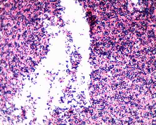


Gram Stain Microbiology Images Photographs


Effect Of The Compound No On Cell Morphology Of E Coli Cells Download Scientific Diagram
View under Microscope (10x, add oil then 100X) Ecoli TSA 7 Heat slide and label 8 Preform Aseptic technique to retrieve bacteria from the slant and smear on slide (remember to add a drop of water) 9 Air Dry Slide 10 Heat Fix slide 11 Preform Gram Stain 12 View under Microscope (10x, add oil then 100X) Mixed TSA and TSB 13The E coli had a Gram Stain reaction color of pink and classified as Gramnegative Both the S epidermidis and B subtilis had a Gram Stain reaction color of purple and then classified as Grampositive (Table 1) Table 1 The Outcome of Gram Stain on Three Species of Bacteria Table 1Escherichia coli Four different strains of Escherichia coli on Endo agar with biochemical slope Glucose fermentation with gas production, urea and H 2 S negative, lactose positive (with exception of strain D "late lactose fermenter";


Staphylococcus Aureus And Ecoli Under Microscope Microscopy Of Gram Positive Cocci And Gram Negative Bacilli Morphology And Microscopic Appearance Of Staphylococcus Aureus And E Coli S Aureus Gram Stain And Colony Morphology On Agar Clinical


Colony Characteristics Of E Coli Sciencing
Jane Buckle PhD, RN, in Clinical Aromatherapy (Third Edition), 15 VancomycinResistant Escherichia coli Escherichia is a gramnegative bacterium, which under the microscope is shaped like a rod with a small tail It is widely distributed in nature (Brooker 08)Escherichia coli (E coli) is part of the normal intestinal flora Some strains are pathogenic and can cause gastroenteritis, UTIThe stain also provided an elimination of Gram () Bacillus cereus and Bacillus subititus, because these bacteria would exhibit rod shapes The orange colony was Gram stained, observed, and recorded Under the light microscope the colony appeared to be pink and purple color, meaning the culture was still mixed
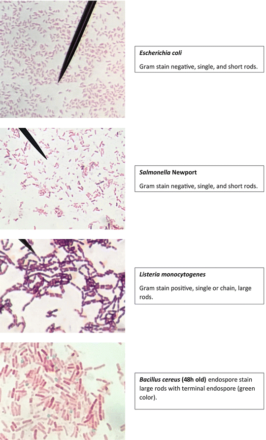


Staining Technology And Bright Field Microscope Use Springerlink
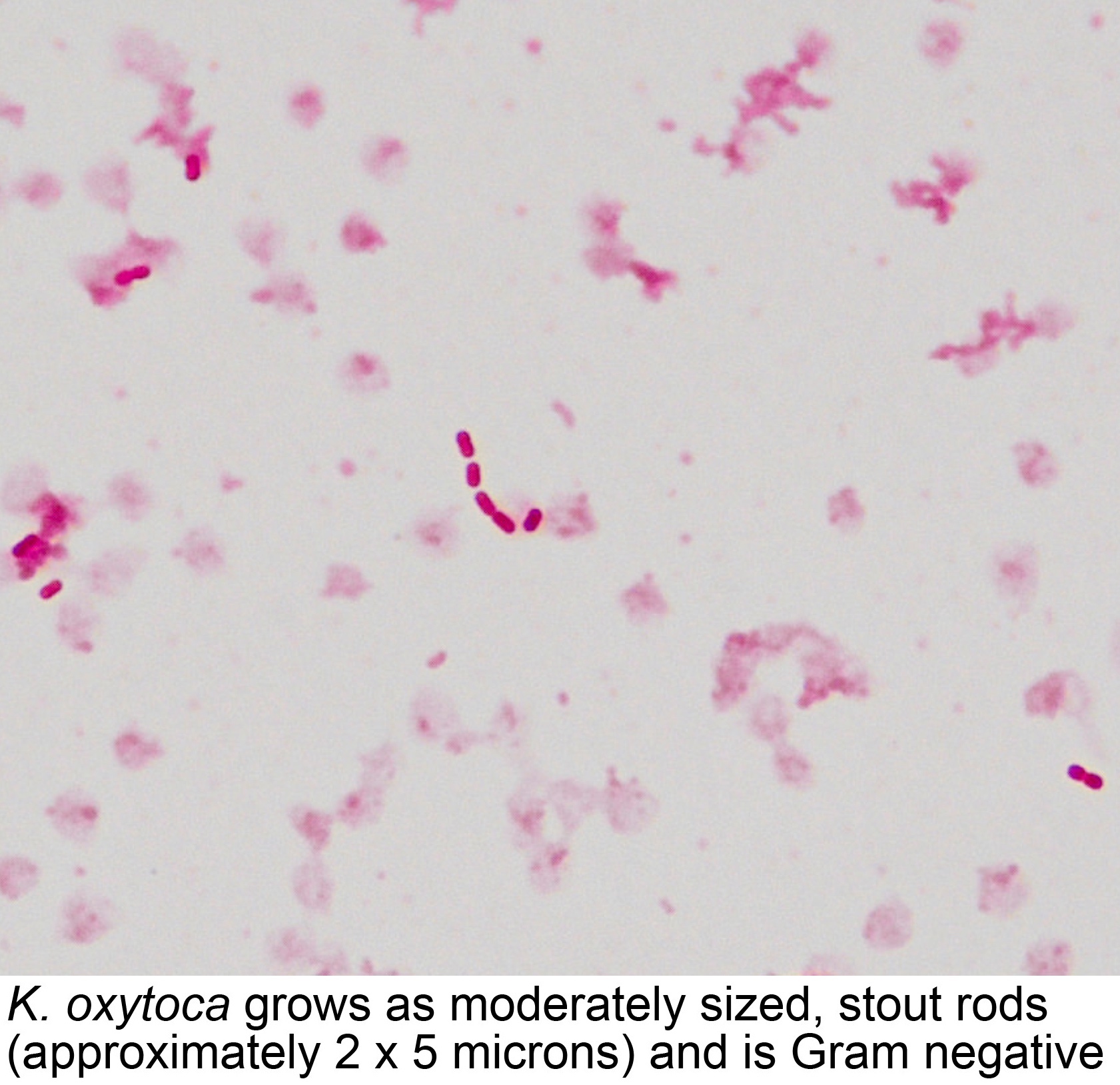


Pathology Outlines Klebsiella Oxytoca


Microscope World Blog Microscopy Gram Staining


Staphylococcus Aureus And Ecoli Under Microscope Microscopy Of Gram Positive Cocci And Gram Negative Bacilli Morphology And Microscopic Appearance Of Staphylococcus Aureus And E Coli S Aureus Gram Stain And Colony Morphology On Agar Clinical


What Does An E Coli Bacteria Look Like Under A Microscope Quora


Pathogenic E Coli
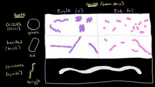


Bacterial Characteristics Gram Staining Video Khan Academy


Microscopy Aquarium Advice Aquarium Forum Community
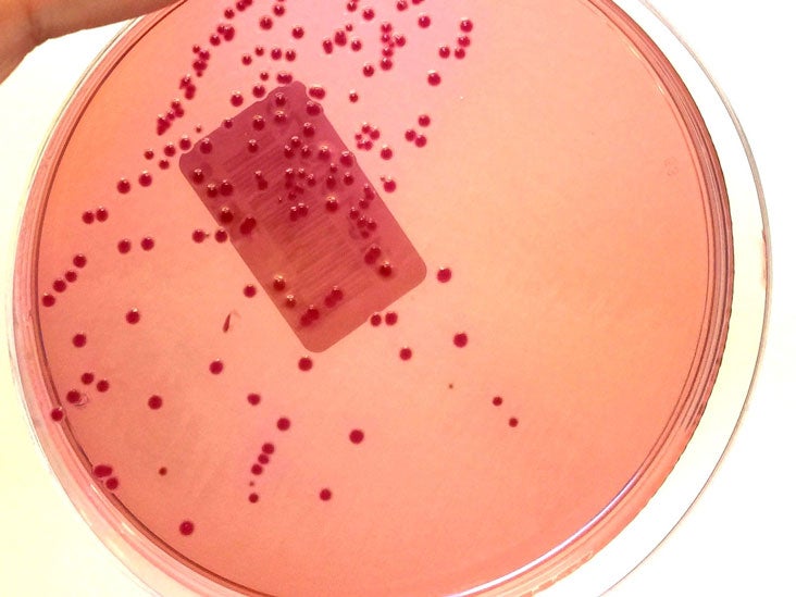


Gram Stain Definition And Patient Education


12 3b Immediate Direct Examination Of Specimen Biology Libretexts


Escherichia Coli Light Microscopy


E Coli Bacteria Britannica


File E Coli Gram Stain Jpg Wikimedia Commons
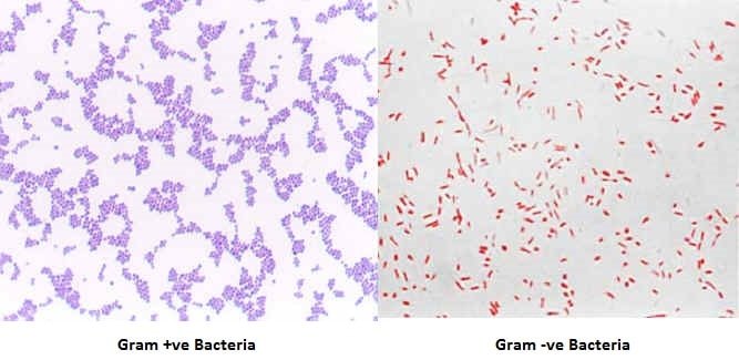


Gram Staining Principle Procedure Interpretation Examples And Animation
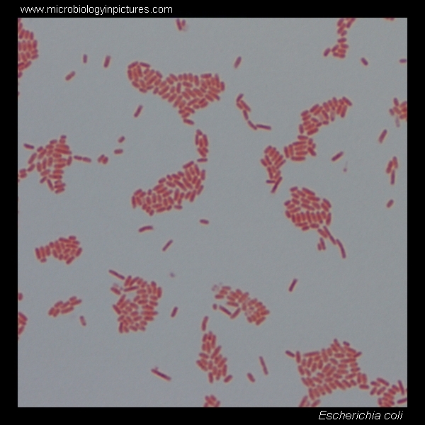


E Coli Gram Stain And Cell Morphology E Coli Micrograph Appearance Under The Microscope E Coli Cell Morphology E Coli Microscopic Picture


A Novel Improved Gram Staining Method Based On The Capillary Tube
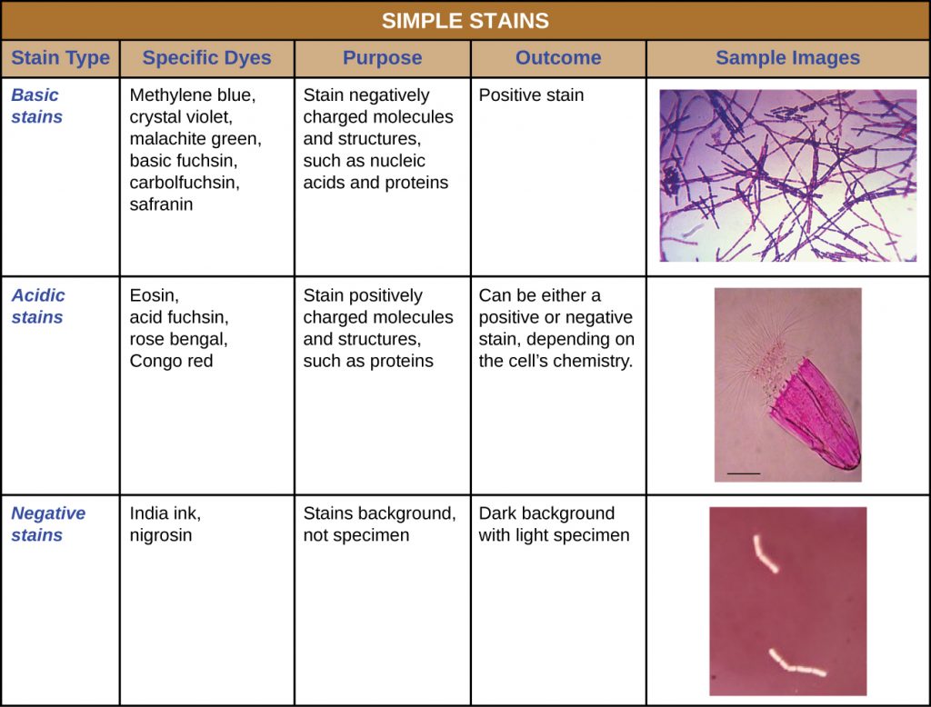


2 4 Staining Microscopic Specimens Microbiology Canadian Edition


Gram Stain


Escherichia Coli Microscope High Resolution Stock Photography And Images Alamy


Sgugenetics Escherichia Coli


Bacteria Under The Microscope E Coli And S Aureus Youtube


Www Mccc Edu Hilkerd Documents Bio1lab3 Exp 4 000 Pdf


Http Coltonanderson1 Weebly Com Uploads 2 4 3 0 Manual Pdf


E Coli Under The Microscope Types Techniques Gram Stain Hanging Drop Method


File Microbiology Gram Stain Jpg Wikimedia Commons


Gram Negative Bacteria Wikipedia


Observing Bacteria Under The Microscope Gram Stain Steps Rs Science


E Coli Bacteria Are Gram Negative Bacilli Or Rods That Inhabit The Bacillus Bacteria Urinary Tract Infection


Observing Bacteria Under The Microscope Gram Stain Steps Rs Science


10pk Escherichia Coli Smear Gram Stain Prepared Microscope Slides 75 X 25mm Classroom Pack 10 Slides In Storage Case Biology Microscopy Eisco Labs Amazon Com Industrial Scientific


Gram Stain Of E Coli Bacterium A Gram Stain Of Shows Gramnegative Download Scientific Diagram


Gram Stain Demonstration Slide 400x 1 Marc Perkins Photography


Pin On Microbiology


Gram Negative Bacteria Under Microscope Page 1 Line 17qq Com


Q Tbn And9gcqkye60ou Johpr02n Mbv1fferrjpdh Lnct7ymdf5qhyia1ld Usqp Cau


Gram Positive And Gram Negative Rods Microscopy Microbiology Microbiology Lab


Escherichia Coli Morphology Mixed Rods Gram Stain Gram Negative Bacilli Growth Characteristics Dubai Khalifa


Escherichia Coli Colony Morphology And Microscopic Appearance Basic Characteristic And Tests For Identification Of E Coli Bacteria Images Of Escherichia Coli Antibiotic Treatment Of E Coli Infections


Optical Microscope Images Of E Coli Cells Following Gram Staining A Download Scientific Diagram


Escherichia Coli Gram Stain Compound Microscope Stock Photo Edit Now


Gram Stain Microbiology Images Photographs


Optical Microscope Images Of E Coli Cells Following Gram Staining A Download Scientific Diagram


Pulsenotes Gram Negative Infections Notes


Q Tbn And9gcqkye60ou Johpr02n Mbv1fferrjpdh Lnct7ymdf5qhyia1ld Usqp Cau


Eiscoprepared Microscope Slide Escherichia Coli Smear Gram Stain Microbiology Fisher Scientific


Escherichia Coli Gram Staining Compound Microscope Stock Photo Edit Now


Microbiology Lab Exercise 10 Smear Preparation Exercise 11 Simple Staining Ex 12 Negative Staining Review Lab Results From Last Week Ex 8 Aseptic Technique Did You Successfully Get Bacteria Growing In Your Broth And Slant Ex 6 The


ᐈ Gram Negative Bacteria Stock Images Royalty Free E Coli Gram Stain Pictures Download On Depositphotos


Www Wiv Isp Be Qml Activities External Quality Rapports Atlas Bacteriology Gram Negative Aerobic And Facultative Rods Pdf
/gram_positive_vs_negative-5b7f26d2c9e77c005746fbd7.jpg)


Gram Positive Vs Gram Negative Bacteria


Bacterial Killing By Dry Metallic Copper Surfaces Applied And Environmental Microbiology


E Coli Gram Staining Page 1 Line 17qq Com


Q Tbn And9gcswouuht13c4cxzkdgvwicdnmwhgkfjwlh40a Eerzzlpwjtmyt Usqp Cau


Escherichia Coli E Coli An Overview Microbe Notes


3a Exercises


Gram Stain Introduction Principle Procedure Result And Interpretation


Gram Stain Wikipedia


E Coli Gram Stain Introduction Principle Procedure And Result Interpret


Staining Microscopic Specimens Microbiology


Laboratory Test 1 Flashcards Chegg Com


Gram Stain Of Escherichia Coli
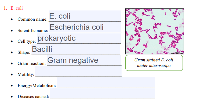


Solved 1 E Coli E Coli Common Name Escherichia Coli S Chegg Com


Biol 230 Lab Manual Gram Stain Of E Coli
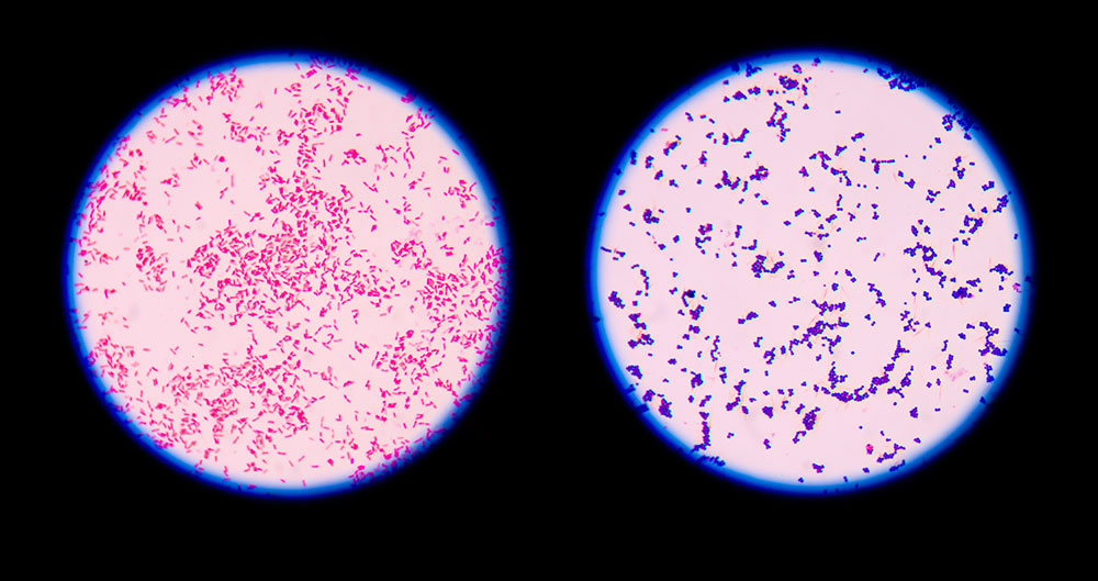


9 Gram Staining Best Practices Microbiologics Blog


E Coli Vs S Aureus Gram Negative Vs Gram Positive Bacteria Youtube


Lab 1


Asmscience Examination Of Gram Stains Of Urine


Gram Stain


Endospore Staining Principle Procedure And Results Learn Microbiology Online


Morphological View Of Lactococcus Culture Under Microscope 100x After Download Scientific Diagram


Gram Positive Bacteria Microbiology


A Long Chain Like Colony Of Streptococcus Bacteria As Seen Under Download Scientific Diagram


Academic Oup Com Labmed Article Pdf 32 7 368 Labmed32 0368 Pdf
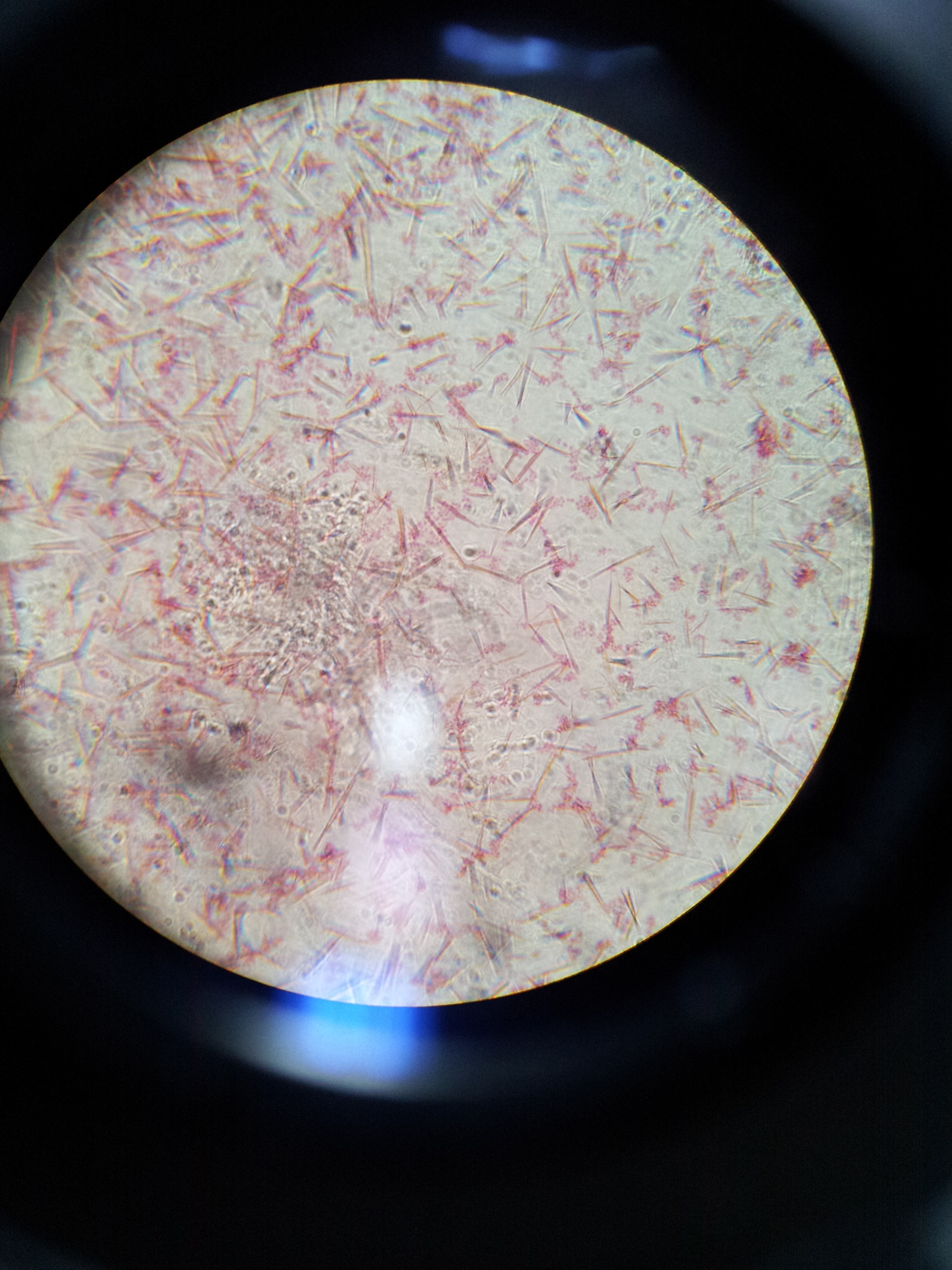


Lab 1 Principles And Use Of Microscope Ibg 102 Lab Reports
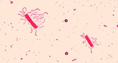


Staining Microscopic Specimens Microbiology


Photographic Print E Coli Bacteria Gram Negative Bacilli Gram Stain Lm X400 By Gladden Willis 24x18in Bacillus Photographic Print Microbiology


コメント
コメントを投稿